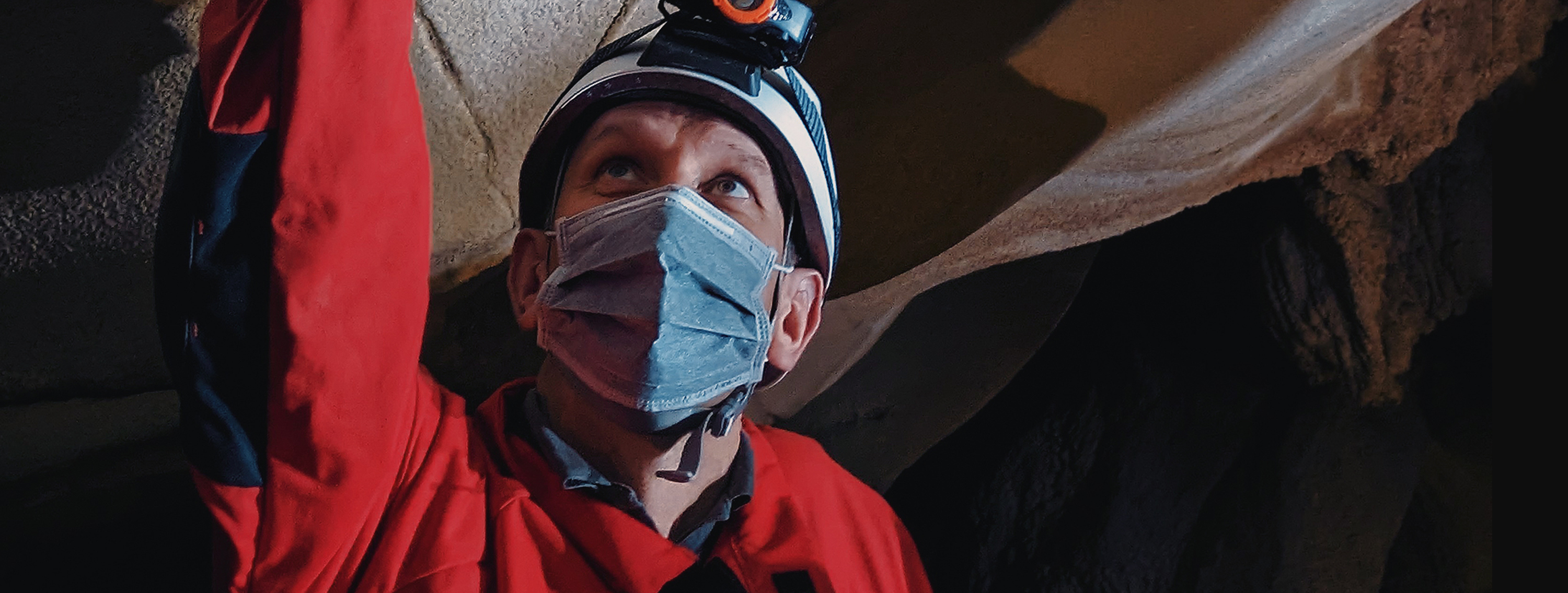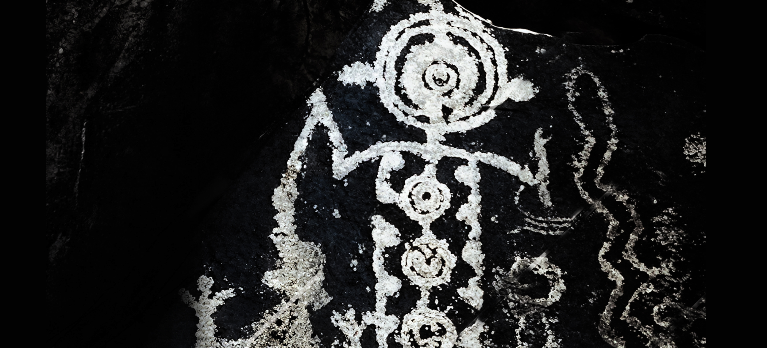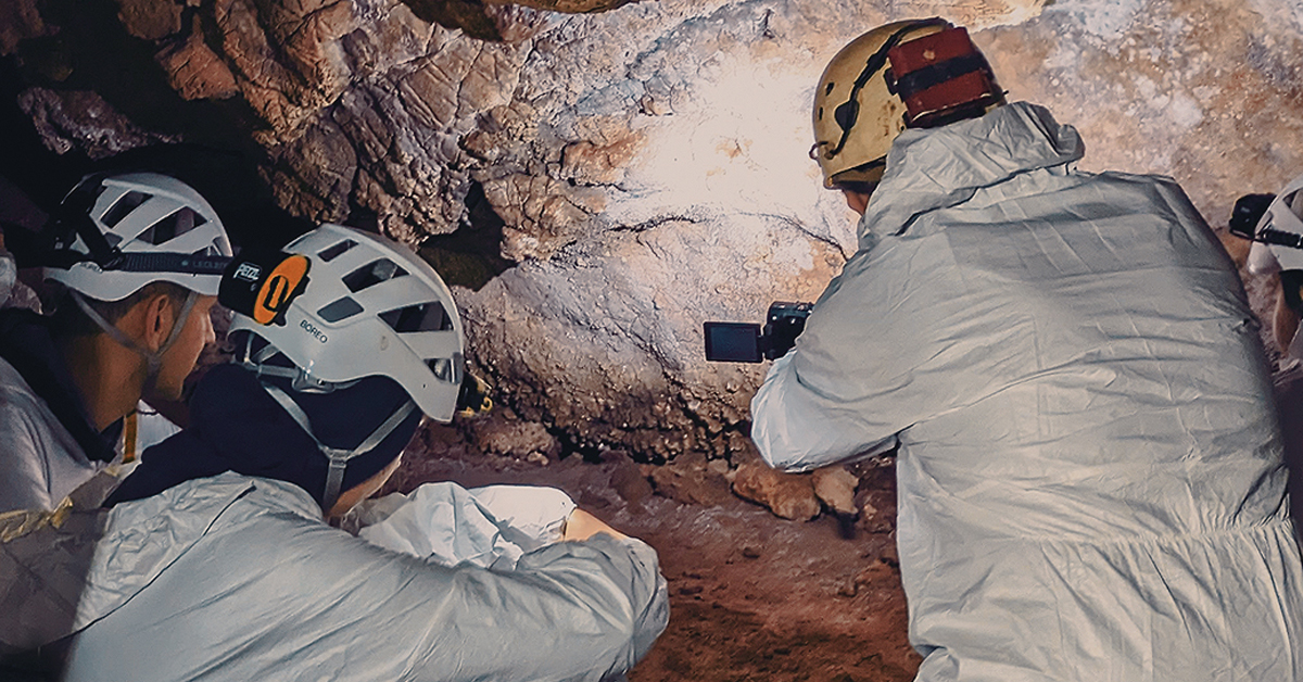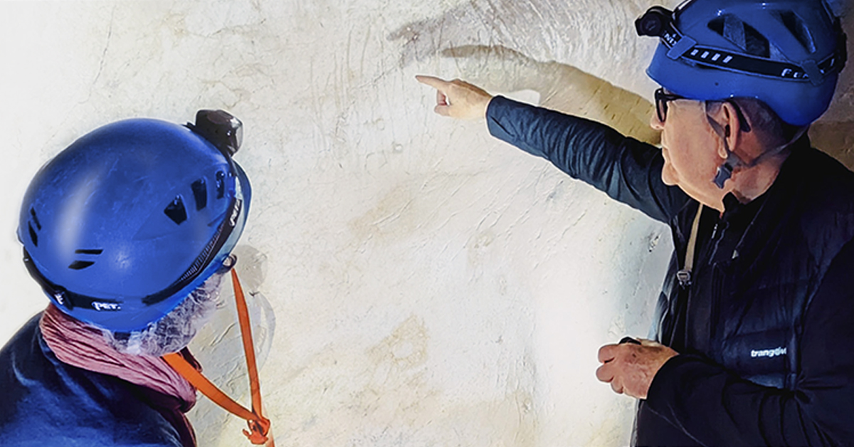

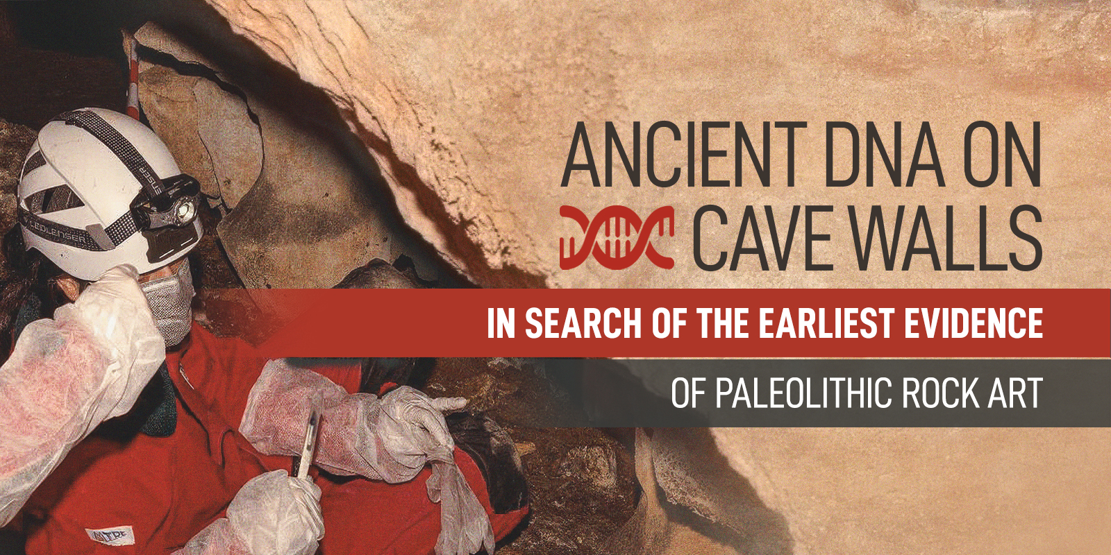
This Project Design outlines the planned expedition to identify rock art panels within several caves located in the Asturias region of Northern Spain. This region, characterised by extensive limestone formations, hosts a significant number of caves that provide evidence of Palaeolithic occupation, with dates spanning from the Neanderthal period (circa 40,000 to 250,000 years ago). Numerous caves in the area have also yielded compelling evidence of Early Modern Human (EMH) presence, dating from approximately 10,000 to 40,000 years ago.
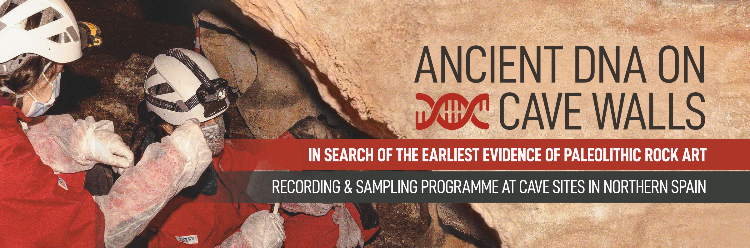
The inaugural "First Art" expedition will focus on La Lloseta Cave, La Loseta 11, and Subore Cave. The objectives of this venture are to conduct geo-prospection for rock art, to document findings through photogrammetric techniques, and to obtain samples for minimum and maximum dating using Uranium-Thorium methods, as well as for the extraction of human and hominin DNA.
First Art, founded in 2018, is composed of a dedicated team of scholars specialising in rock art science research from early prehistoric contexts. The team has previously managed projects in Israel, Portugal, Spain, South Africa, and Wales. As part of our fieldwork initiative, we collaborate closely with the DNA team from the Max Planck Institute in Leipzig, Germany and dating specialists from the University of Southampton, UK, and Nanjing Normal University, China.
The following sections of this Project Design provide an overview of the historical context of our work program (Section 2), the scientific methodologies employed (Section 3), the project's objectives (Section 4), the team and its responsibilities (Section 5), and the expected methodologies and outcomes (Section 6). This document is supported by a detailed bibliography.
For the duration of this proposed project, all participants are covered by accidental insurance administered through their respective university institutions.
For many years, it was believed that Palaeolithic rock art was an exclusive product of Homo sapiens as it was thought to be a cultural product and as such, it required high levels of cognitive ability, including symbolic behavior, abstract thought and, possibly, a complex language or sign system. At this time, these attributes were considered unique to Homo sapiens. This led to the assumption that only Sapiens could create art. However, with new scientific techniques, and recent uncorrupted discoveries, using Uranium–thorium dating methods for sampling calcium carbonate crusts covering prehistoric paintings, the possibility of Neanderthal involvement in cave art has recently been proposed and thus challenges long-held assumptions. Essentially, this debate centers around the question of whether Neanderthals, an extinct archaic species of human, had the cognitive and creative abilities to produce art or some form of pictorial language system. Based on crucial dating ranges, current debate exists on whether the art represents Homo sapiens, or possibly, Neanderthal artistic endeavor, or simply is it the result of natural processes. Our novel strategy is to expand the number of sampled caves, resulting in the analysis of a large corpus of pigment samples through physical/chemical analysis, chronologically framing them using Uranium–thorium dating techniques and to validate our sampling programme using two laboratories.
Running parallel to this sampling programme will be the search for the presence of human DNA in pigments. This protocol will help to answer the fundamental questions of who created the art and determine its age. We propose that archaic hominids may have had the brain capacity to express themselves through the medium of art. FIRST ART believes we are on the brink of a new scientific initiative to unravel the mysteries of pigments and the contexts in which they are embellished. We suggest that previous thinking about our ancestors requires an overhaul.
The research of Palaeolithic cave art has gained prominence since the beginning of the last century and has continuously evolved to the present day, with significant milestones marking important advancements in each stage. In this regard, during the first half of the 20th century, the studies conducted by Henri Breuil stood out, alongside the invaluable contributions of researchers such as Hugo Obermaier, Émile Cartailhac, Louis Capitan, Denis Peyrony, Lorenzo Sierra, Marcelino Sanz de Sautuola, Eduardo Hernández Pacheco, and Hermilio Alcalde del Río (Moure, 1999). These studies were conducted within the framework of the development of prehistory as a scientific discipline and allowed for the establishment of the initial diachronic sequence for cave art based on technical and stylistic criteria (Breuil, 1952).
The second half of the 20th century is mostly characterized by the development of structuralism and its application to the interpretation of prehistoric art, led by André Leroi-Gourhan (1965) and Anette Laming-Emperaire (1962). Their work was based on Max Raphael's book "Prehistoric Cave Paintings" (1945). They conceptualized prehistoric expressions to graphically represent the worldview of these ancient societies through a symbolic language, in which figures were recurrently organized within the physical space of the cave. The cave was thus understood as an "organized sanctuary" where the two major opposing principles—masculine versus feminine, bison versus horses—were arranged, along with their associated symbols, which appeared repeatedly in different areas of the cave.
Despite the inconsistency of the structuralist proposal in relation to an archaeological reality that increasingly questioned the foundations supporting such interpretation, this theory remained popular until the early 1990s. It was during this time that Jean Clottes and David Lewis-Williams (1996) highlighted shamanism as the latest major interpretative proposal for Paleolithic art. However, around the same period, there was also a re-evaluation of studies on prehistoric art, questioning the validity of the prevailing stylistic evolution. This new debate, based on the emergence of new direct dating methods in prehistoric art research (such as C14 AMS), even created a new term: "the post-Stylistic era" (Lorblanchet and Bahn, 1993). Despite the immediate publication of results obtained in Chauvet cave (Clottes et al., 1995), this term faced refutation by a significant group of researchers who were critical of the validity and appropriateness of C14 AMS for directly dating cave art. These researchers promoted for maintaining the use of style as the basis for determining the evolution of Paleolithic figurative art (Züchner, 1999; 2007).
This preamble immerses us fully into the 21st century, where we witness the extraordinary diffusion and utilization of new documentation systems primarily based on the advancement of digital photography. The potential of specialized software has significantly contributed to the processing and enhancement of visualizing these digital images (Harman, 2005), while the integration of support as a referential element in the analysis of rock art representations has been facilitated using high-precision 3D scanners (Angás, 2019). This technological progress has not only led to the revision of some of the major assemblages of European Paleolithic cave art (Collado and García, 2022; Jouteau et al., 2020; Balbín et al., 2022) but has also resulted in the discovery of new sites and the expansion of Paleolithic cave art into territories that had previously remained outside the purview of this phenomenon (Fernández et al., 2021).
However, despite all these new technological contributions and the emergence of new research approaches that recurrently emphasize the interaction between support and cave art (Petrognani et al., 2015; Ferrier et al., 2017; Jouteau et al., 2019), one paradigm remained unchanged throughout these nearly 150 years of archaeological research. The European Homo sapiens had been considered the only species capable of using rock surfaces to produce cave art, thus restricting the timeframe of its emergence to the early phases of this species' presence in southern Europe, around 40,000-35,000 years BP. This perspective failed to acknowledge that, even within the realm of portable objects, there is evidence that the earliest graphic representations date back over half a million years and, of course, extend well beyond the strictly European context (Joordens et al., 2015; Collado, 2012).
In this regard, towards the end of the first decade of the 21st century, the methodology of Uranium series dating emerged to determine the age of Paleolithic cave art, and its initial results indicated a significantly older age for part of the Paleolithic iconographic repertoire (Pike et al., 2012). The scientific community reacted in a similar way as they had in the 1990s when C14 AMS dating was introduced, generally adopting a critical stance, often expressing outright opposition, towards both the methodology employed and the new chronological evidence it provided. Specifically, aspects were questioned regarding the reliability of chronological measurements using the Uranium series on the samples, whether the Uranium series method was applied correctly, the accuracy of stratigraphic relationships between the imagery and the samples, and whether the dated pigment was a result of intentional human action (Clottes, 2012; Pons-Branchu et al., 2014; Sauvet et al., 2015). Naturally, the supporters of this dating method responded to these criticisms (Pike et al., 2017) and, despite the controversy, continued to present new dating that consistently resulted in older dates for cave art (Hoffman et al., 2016). Furthermore, this line of research was enriched by new evidence emerging from other research groups in support of the aging of cave art dates (Rodríguez-Vidal et al., 2014). In this context, a turning point occurred in 2018 when the chronological results obtained from Uranium series dating of calcite crusts covering cave wall figures in three Spanish caves—La Pasiega, Maltravieso, and Ardales—were published. These results confirmed the authorship of these cave art by individuals of the Neanderthal species (Hoffman et al., 2018). A prompt response followed (White et al., 2019), sparking a fascinating scientific debate mainly focused on acknowledging the validity of the method used to date the crusts (Slimak et al., 2018; Aubert et al., 2018; Hoffman et al., 2018b; 2020; Pons-Branchu et al., 2020).
Considering these circumstances, we consider ourselves to be engaged in a crucial debate for understanding the origins of Paleolithic cave art, which, however, is still far from being solved mainly because essential issues have been overlooked. First and foremost, it is important to highlight the extremely limited representativeness of the number of conducted projects. As an example, in the Iberian Peninsula, where we are aware of more than 600 sites with Paleolithic cave art, only 269 datings had been undertken as of 2021 (117 using C14 AMS, 137 using Uranium series, and 15 using Thermoluminescence -TL-) (Ochoa et al., 2021), corresponding to only 36 caves. This means that in one of the key territories for understanding the origin and evolution of cave art, only 6% of the sites with Paleolithic cave art had been dated, and among all these samples, less than 1% corresponded to figures with an age exceeding 30,000 years. However, it is not only a matter of constraining the deficiencies to strictly chronological aspects. In fact, to this day, there is no comprehensive iconographic catalogue of the earliest cave art expressions, and similarly, the technical and stylistic criteria that influenced the making process of the most archaic imagery have not been established. All of this prevents us from approaching the study of the initial stages of cave art in a comprehensive manner, making it impossible to truly understand the territorial extent and the way in which this phenomenon spread. Moreover, it is far from determining which human species or species were involved in this process of graphic creation and how it influenced the evolution of their cognitive abilities.
With the challenges laid out, the team spearheading the First Art project has embarked on a significant mission: to unravel the origins of cave art by comprehending the symbolic repertoire that humans, irrespective of their species, were capable of depicting on rocky surfaces. To achieve this, a methodology has been designed to provide a clear and definitive response to the stated objective. To accomplish this, our work will be based on the combined action of three complementary processes:
- Establishing a consistent and accurate chronological framework within which the earliest graphic expressions took place.
- The characterization, based on technical, stylistic, and morphological criteria, of those graphic elements that dating studies associated with the emergence of art. The objective is to establish the iconographic repertoire of the earliest symbols that humans, regardless of their species, could represent on a wall, as well as the technical procedures involved in the operative chain to make it and the materials used. In this sense, while recognizing that the clearest evidence of this process is to be found in southern Europe and more specifically in the Iberian Peninsula, where we will focus our efforts as a priority; we want to overcome the Eurocentrism in which we recurrently fall into and extend the research globally to all those places where parietal cave paintings have been documented that are more than 40,000 years old (Brumm et al., 2021). Once this figurative and technical corpus has been defined, we will be able to carry out wideranging evaluations of the development of this symbolic process, such as, for example, establishing spatial analyses that clarify how the expansion of this first symbolic universe took place, what its core areas were, if any, or whether, on the contrary, if it was a multifocal process generated in different territories and how they were related to each other.
- The identification of the human groups that played a pivotal role in the genesis of the earliest graphic representations by examining traces of human DNA present in pigment samples derived from the figures comprising the iconographic repertoire associated with the initial phases of cave art, as determined by the chronological, technical, and stylistic criteria outlined in the preceding sections. This research aims not only to ascertain which species or individuals were responsible for painting or engraving these symbols on cave walls but also to leverage the full potential offered by contemporary DNA studies. By doing so, it seeks to shed light on the involvement of men and women in the early stages of the creative process, discern the age groups implicated therein, and uncover patterns of movement or areas of influence by establishing genetic connections between these symbols and specific human populations.
The three processes outlined above form the fundamental pillars of the FIRST ART project. However, they are not independent actions, but rather integrated and interdependent components that work together. The outcomes derived from each process complement one another, contributing to a unified and cohesive response to the challenges at hand.
In this regard, the establishment of a chronological framework constitutes a cornerstone of paramount importance. While we acknowledge the potential of recent trends such as fuliginochronology (Medina et al., 2023; Vandevelde et al., 2018) or thermoluminescence (Bonneau, 2016) for achieving specific chronological contexts, our leading reliance will be on the Uranium Series method to determine the temporal range associated with the origins of art. The choice is based on the effectiveness and reliability of the employed method (Hoffmann et al., 2016b). However, we aim to enhance it further by incorporating geological studies to properly characterize the carbonate samples that will be dated, thus avoiding issues arising from diagenetic processes or open-system behavior (Pons-Branchu et al., 2020). Additionally, whenever possible, the Uranium Series method will be complemented by the C14AMS method. Nonetheless, it is important to address the main limitations that arise from the latter for our research. These include the inability to date motifs created with inorganic pigments or engravings, and most significantly, surpassing the barrier around 50,000 years, which represents the maximum age that can be achieved through C14AMS dating of organic pigments. We anticipate that a significant portion of the representations included in this initial catalogue will far exceed this temporal limit. In addition, whenever sample size allows, it is a priority objective to split the sample and process it in several different laboratories under similar protocols. Finally, the potential to document human DNA in the pigment of some of the figures that have been dated will provide an unparalleled control protocol, as it will allow us to cross-check the dates provided by the dating method with those indicated by the results of the DNA identified in the pigment (Gansauge et al., 2014; Braadbaart et al., 2020; Zavala et al., 2022; Hessel et al., 2023).
The determination of this chronological framework allows us to confidently establish the first iconographic corpus of engraved and painted representations within this chronological period, as well as the materials used in their creation and the technical procedures involved in their implementation. In the pursuit of these objectives, physicochemical methodologies for pigment and binder characterization, such as Scanning Electron Microscopy combined with Energy-Dispersive X-ray Spectroscopy (SEM-EDX), Fourier Transform Infrared Spectroscopy (FTIR), and micro-Raman spectroscopy (μ-Raman), will play an important role in the FIRST ART project. These analyses will be carried out by the laboratories at the Polytechnic Institute of Tomar (Portugal) and the University of Ferrara (Italy), which, as part of the FIRST ART project, will be responsible for precisely determining the techniques and preparation methods used by these ancient groups for their pigments. These analyses will identify the existence of authentic "color palettes" that can be recognized in various territories. Traceological analyses (Zotkina and Miklashevich, 2016; Rivero and Gárate, 2020) will also be extensively conducted, as well as three-dimensional documentation of both the figures and the rocky-support surfaces. This will be achieved using high-resolution structured light scanners and photogrammetric compositions configured with SIFT-based software (Angás et al., 2015; Angás, 2019; González-Aguilera et al., 2009; Richardson et al., 2013; Feruglio et al., 2013).
Once the chronological framework is established and the iconographic and technical aspects of the early cave art are defined, the third major part of the FIRST ART project, and one of its most innovative contributions, will be to leverage a large number of pigment samples available to search for evidence of human DNA. To date, human DNA has been successfully identified in sediments associated with settlements (Rohland et al., 2018) and even in personal adornments (Essel, 2023), but the examination of pigments from cave art figures has never been achieved before. Undoubtedly, the processing that these pigments underwent during their preparation (mixed with hands and possibly with their own organic fluids - saliva, urine) or their application (sometimes blown after being held in the mouth) raises hopes that traces of hominid DNA may be preserved in some of the pigment samples that will be analyzed. In this regard, the extensive expertise and experience in ancient DNA sequencing techniques owned by the team at the Max Planck Institute for Evolutionary Anthropology, who will be responsible for this task within the FIRST ART project, give us great hope that we can successfully accomplish this endeavor. This achievement would undoubtedly mark a turning point in the interpretation of prehistoric art, as we would obtain irrefutable data that will allow us to address controversial topics such as the role that men or women played in the creation of these graphics (Adovasio et al., 2007; Snow, 2013), the presence of children associated with the cave representations, and even, in the case of obtaining a significant evidence, the possibility to analyze patterns of movement or diffusion of genetically related human groups, as well as the symbols they created and broadcasted during their migrations. Additionally, identifying this DNA would provide us with a precise chronological framework for the analyzed sample, which in turn could serve as an excellent validation system for other chronological methods employed in determining its age.
The implementation of all the actions described so far would, in and of itself, constitute a fundamental contribution to the intrinsic understanding and dissemination of the origins of cave art. However, in the FIRST ART project, we have aimed to go a step further by subjecting this entire symbolic corpus to a critical analysis from the field of cognitive neuroscience, to understand the influence that these early rock art expressions may have had on the brain activity of these ancient hominids (Perote and Martín-Loeches, 2012). To achieve this, the team responsible for the Cognitive Neuroscience Section at the Joint UCM-IS CIII Center for Evolution and Human Behavior, will undertake this task during the project's development. They will design a series of experiments aimed at determining the significance of the creation and use of these symbols in the development of symbolic capacity in fossil hominids, and how it may have influenced the evolution of other key cognitive abilities such as technological capacity, consciousness, and knowledge sharing. Ultimately, this research aims to uncover the extent and way the creation and dissemination of early artistic expressions shaped the evolution of human behavior.
The FIRST ART project has embarked on a significant mission: to unravel the origins of prehistoric art by comprehending the symbolic repertoire that humans, irrespective of their species, could depict on rocky surfaces. In essence, we aim to uncover the origins of prehistoric art. This is what sets us apart from any other project, as we want to venture into unexplored territory while maintaining the utmost respect for the theories that have brought us here. At the same time, we want to free ourselves from preconceived ideas that could limit our investigative creativity and results. To accomplish this, our work will be based on the combined action of three complementary processes:
- To establish a consistent and accurate chronological framework within which the earliest graphic expressions took place.
- To characterize, based on technical, stylistic, and morphological criteria, the graphic elements that dating studies associate with the emergence of art. The objective is to establish the iconographic repertoire of the earliest symbols that humans, regardless of their species, could represent on a wall, as well as the technical procedures involved in the operative chain to make it and the materials used. In this sense, while recognizing that the clearest evidence of this process is to be found in southern Europe and more specifically in the Iberian Peninsula, where we will focus our efforts as a priority, we want to overcome the Eurocentrism in which we recurrently fall into and extend the research globally to all those places where parietal cave paintings have been documented that are more than 40,000 years old (Brumm et al., 2021). Once this figurative and technical corpus has been defined, we will be able to carry out wide-ranging evaluations of the development of this symbolic process, such as, for example, establishing spatial analyses that clarify how the expansion of this first symbolic universe took place, what its core areas were, if any, or whether, on the contrary, if it was a multifocal process generated in different territories and how they were related to each other. To achieve this, we will formulate a scientific process that hopefully will stand against constructive criticism.
- To identify the human groups that played a crucial role in the genesis of the earliest graphic representations by examining traces of human DNA present in pigment samples derived from the figures comprising the iconographic repertoire associated with the initial phases of cave art, as determined by the chronological, technical, and stylistic criteria outlined in the preceding sections. This research aims not only to ascertain which species or individuals were responsible for painting or engraving these symbols on cave walls but also to leverage the full potential offered by contemporary DNA studies. By doing so, it seeks to shed light on the involvement of men and women in the early stages of the creative process, discern the age groups implicated therein, and uncover patterns of movement or areas of influence by establishing genetic connections between these symbols and specific human populations.
The acquisition of a comprehensive understanding and widespread dissemination of the origins of cave art would already constitute a significant contribution to intrinsic knowledge. However, the FIRST ART project aims to transcend conventional boundaries by embarking on a new and ambitious endeavor. In addition to the realms of archaeology and art, we intend to delve into uncharted territory by subjecting the symbolic corpus we have meticulously defined to a critical analysis rooted in cognitive neuroscience. Our objective is to elucidate the potential impact of these early prehistoric art expressions on the brain activity of archaic hominids (e.g., Perote and Martín-Loeches, 2012). Essentially, we seek to comprehend the extent and way the creation and diffusion of the first artistic expressions influenced the evolution of human symbolic behavior. By merging archaeological research with insights from cognitive neuroscience, we aim to shed light on the profound cognitive and behavioral transformations engendered by the earliest forms of artistic expression.
The success of the FIRST ART project's objectives relies on the establishment of clear and precise goals, along with well-defined strategies, and the involvement of a competent and proficient technical team to ensure its successful execution. Therefore, the professionals who have dedicated themselves to carrying out the diverse actions throughout the project's duration, as substantiated by the provided letters of support, embody not only a longstanding history of collaboration but also possess acknowledged technical expertise and extensive research experience.
The planned analytical procedures in the areas of ancient DNA identification/sequencing in pigments will be conducted by the expert team at the Max Planck Institute for Evolutionary Anthropology in Leipzig, Germany. The University of Ferrara in Italy and the Polytechnic Institute of Tomar in Portugal will collaborate to develop the physicochemical methodologies (including FTIR, RAMAN, SEM-EDX and XRF) for pigment and binder characterization, as well as the determination of the geological composition of the dated samples.
The Cognitive Neuroscience Section of the UCM-IS CIII Joint Center for Evolution and Human Behaviour will lead the neurocognitive experiments aimed at understanding the significance of the creation and utilization of the earliest representations of parietal cave art in the development of symbolic capacity in hominids. The coordination of all participating teams, including the Max Planck Institute for Evolutionary Anthropology, the University of Ferrara, and the Polytechnic Institute of Tomar, will be carried out by the project's Principal Investigator (PI) Hipólito Collado Giraldo. The PI will also directly supervise the sampling and three-dimensional graphic cataloguing campaigns conducted at the selected cave art sites for research purposes.
The methodological strategy designed for the FIRST ART project is structured into the following work phases:
Phase 1
Site selection and Panel identification of specific panels and figures.
Documentary record and Sampling. A comprehensive documentary record is created using 2D tracings, high-resolution digital photography, and 3D scanning techniques.
Phase 3
Laboratory Analysis of samples. The samples obtained during the initial sampling process undergo laboratory analysis. This analysis encompasses various aspects, including chronology determination, pigment characterization, and DNA examination. These analyses will yield initial results that contribute to the overall understanding of the studied rock art.
Phase 4
Repeat Sampling and Laboratory analysis: The fourth phase of the methodology involves a second sampling process and the repetition of laboratory analysis (i.e., dual sampling strategy). This step is specifically targeted towards rock art motifs that have shown potential indications of antiquity or have provided evidence of human DNA in the pigment during the initial analyses. The primary objective of this phase is to verify and validate the results obtained from the initial sampling process. By repeating the sampling and analysis, we aim to ensure the accuracy and reliability of the data, either confirming or discarding the initial findings. This rigorous approach contributes to the robustness and credibility of the research outcomes within the FIRST ART project.
Phase 5
Development of Neurological Experiments and Dissemination of Results. Neurological experiments will be developed using the compiled catalogue of figures from previous documentation and analysis work. Results will be disseminated and published.
6.1. Phase 1: Site selection and Panel identification of specific panels and figures
It is proposed as a collaborative work with the researchers responsible for each of the rock art sites. The FIRST ART project does not aim to initiate new research projects in each of the caves and rock shelters with rock art that will be studied, but rather to integrate into the ongoing research projects by establishing a collaboration which FIRST ART will benefit from the expert and intrinsic knowledge that the researchers responsible for these sites have. In return, FIRST ART will provide them with a wide range of tools and analytical capabilities that can be a great opportunity to enhance the knowledge of these sites. This collaborative strategy streamlines administrative procedures by integrating with authorized research teams, eliminating the need for additional documentation and sampling authorizations.
This initial visual recognition will allow for the assessment of the conservation status of the panels and figures, their technical characteristics, and the presence and viability of elements that can be analyzed (underlying or overlapping calcite crusts on the motifs, materials suitable for analysis or dating near the panel, potential contamination factors, etc.). A portable digital microscope will be used to determine areas of the figure with higher pigment content and to ensure the stratigraphic relationship between the paint or engraving and the calcite crusts. Similarly, priority will be given to sampling processes in the peripheral areas of the figure, as well as remnants of pigment that may have been preserved on the panel.
6.2. Phase 2. Documentary record and Sampling
6.2.1. 2D and 3D graphic recording of rock art figures and panels
The workflow implemented for conducting the 2D/3D graphic documentation of each chosen assemblage of rock art figures has been meticulously developed with considerations for noninvasiveness, precise data acquisition, and efficient resource utilization. This has been achieved by employing economically accessible instrumentation and optimizing the process to ensure the highest level of data accuracy.
To accomplish the 3D documentation, we will leverage the capabilities of LIDAR (Light Detection and Ranging) sensors, which have been integrated into modern mobile devices such as the iPhone 14 Pro. This will be complemented by the advanced TrueDepth camera system. Together, these technologies enable the generation of exceptionally accurate 3D models in near real-time, even in challenging light conditions, utilizing wavelengths of approximately 1500 nanometers.
During this process, a low-power pulsed light beam is directed onto the surface of the panel. The purpose of this is to measure the distance between the rock surface containing the rock art representations and the mobile device. This measurement enables the creation of a highly detailed three-dimensional model that accurately captures the intricacies of the documented graphic space. The processing and texturing of the generated meshes will be performed using the Scaniverse software (https://scaniverse.com/). This software offers comprehensive editing and sharing capabilities for 3D content directly from the scanning device. Its use allows for real-time visualization of the three-dimensional files, enabling on-site verification within the cave, and ensuring the accuracy of the captured data and sufficient coverage of the scanned area. Finally, the resulting models will be exported in OBJ and FBX formats, which are widely compatible with various 3D modeling software.
Simultaneously, a thorough 2D documentation process will be carried out for both the panel and the selected rock art motifs. A series of high-resolution photos in RAW format will be captured using calibrated lenses of 24mm, 50mm, and 100mm, which will be mounted on a Canon 6D Mark II camera featuring a 26.2 MP sensor. To ensure stability and precise image capture, the camera will be supported by a three-axis stabilizer system (Ronin DJI RCS2) mounted on a tripod. Additionally, remote, and delayed shooting systems will be employed to optimize the photography process.
To ensure the highest level of detail in the photographic recording, a proximity to the panel will be maintained, despite the resulting shallow depth of field. To address this challenge, the focus stacking macro photography technique will be employed. This technique involves capturing a series of photos, each with a different focal point, and then merging them to create a single image that encompasses all the focus points obtained. To capture the entire panel, aligned photographic sweeps, either vertical or horizontal, will be conducted with a minimum overlap of 30% between each photo. To maintain consistency in brightness, contrast and white balance across all photographic recordings, radiometric adjustments will be applied. A standardized and calibrated color chart, such as the X-Rite ColourChecker Passport, will be included in each shot. This will enable the correction of light temperature in a homogeneous manner for each image, ensuring accurate color representation.<\p>
Following the photographic documentation, all images will undergo processing using Structure from Motion (SfM) software. This software will analyse the images and generate threedimensional point clouds and high-resolution orthophotos of the detailed figures and the panel. To illuminate the rock art representations during the graphic recording processes, LED cold light spotlights will be exclusively utilized. These spotlights have been specifically chosen to ensure that the illumination is completely harmless to the rock art (in terms of heat radiation), preserving its integrity and minimizing any potential damage.
6.3. Sampling processes
The implementation of this activity holds utmost importance within the framework of the FIRST ART project, as it is through these three types of analyses (chronological, component characterization, and DNA presence determination) that we can establish the key evidence required to identify the graphemes associated with the emergence of parietal cave art. Therefore, we have designed a meticulous protocol for sample collection that also complies with the requirements and guidelines outlined in the "Code of Ethics for the Practice of Sample Collection Developed by the American Institute for Conservation" (A.I.C. 2015).
Step A:
The initial stage involves identifying the extraction area and performing a thorough visual examination, utilizing a portable microscope whenever feasible. This examination aims to gather detailed stratigraphic information about the sample in relation to the cave art, assess its cleanliness, consistency, dryness, color, and determine the material type (such as calcite crust, pigment, charcoal, clay, etc.). Additionally, potential contamination factors are identified during this inspection. To ensure systematic data collection, all relevant information, including the precise sample location, topographic details of the panel within the cave, and pre- and postextraction photographic documentation, will be recorded in a standardized format.
Step B:
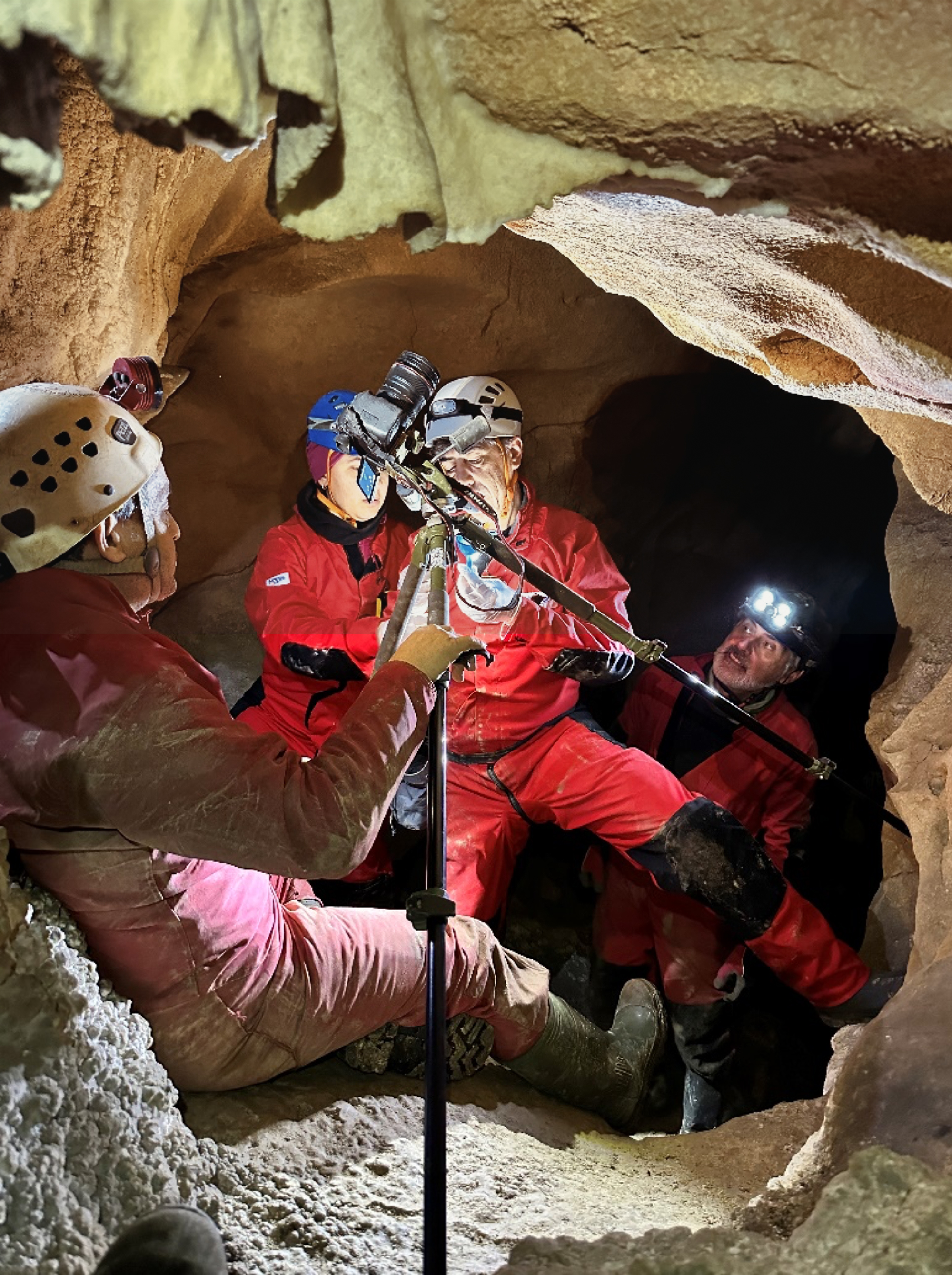
The sample extraction procedure is conducted during this stage. This process will always be performed mechanically, using a variety of equipment depending on the characteristics and hardness of the sample, such as tungsten scalpels or microdrills with tungsten bits or diamond cutting discs. All tools used must undergo a rigorous sterilization process, and their active parts (scalpel blades, bits, or discs) should only be used for a single sample. Immediately before the extraction, the sampling area must be cleaned thoroughly to prevent potential contamination from dirt. The sample will be collected directly into a sealed test tube (Eppendorf type), which will be held by an assistant. Both the sampling technician and the assistant must wear nitrile gloves and face masks to avoid contaminating the samples. The sample size will depend on the type of analysis to be performed and variable factors such as uranium concentration, age range, and even the instrumentation used. In any case, the sampling ranges established in FIRST ART will range from 2 to 5 mg for pigment or charcoal samples and from 5 to 10 mg for calcium carbonate or sediment samples. Whenever possible, larger samples will be collected.
Once detached from the rock surface and securely placed inside a sealed test tube, the sample will be properly identified using a labelling system that includes three letters identifying the cave art site, followed by a serial number separated by a hyphen (for example, for the Maltravieso Cave, the label would include these data: MAL-001). This identification will be referenced in the sampling form, and the tube will be sealed and stored under appropriate humidity and temperature conditions.
6.4. Phase 3. Laboratory analysis of samples
After obtaining the samples from each of the selected panels and figures for our study, they will be sent to their respective laboratories to obtain the corresponding chronological results, identify their components, and potentially confirm the presence of DNA in the sampled pigments.
6.4.1. Chronological Analysis. Uranium Series
While remaining attentive to the latest advancements in obtaining precise chronological frameworks, such as fuliginochronology (Medina et al., 2023; Vandevelde et al., 2018) or thermoluminescence (Bonneau, 2016), our primary approach for establishing the temporal range associated with the origins of art will be the Uranium Series method. We have chosen this method due to its effectiveness and reliability (Hoffmann et al., 2016), although we aim to enhance its accuracy by conducting thorough geological studies to properly characterize the carbonate samples intended for dating. This approach will help mitigate potential issues related to diagenetic processes or open systems (Pons-Branchu et al., 2020). Additionally, when feasible, we will supplement our research with the C14AMS method, addressing the main date range limitations that arise in our specific context. The U/Th methodology has emerged as a crucial technique in geochronology, finding extensive applications in various fields. It has been successfully utilized in reconstructing sea-level variations using corals (Bard et al., 1996; Fairbanks, 1989), calibrating the radiocarbon time scale (Fairbanks et al., 2005), and dating carbonates derived from stalagmites, travertines, and corals (Adkins et al., 1998; Frank et al., 2006; Mallick and Frank, 2002). Its versatility and reliability make it a valuable tool in accurately determining the ages of geological and archaeological materials.
The methodologies used in laboratories for U/Th dating are relatively standardized and documented in several studies (Shao et al., 2017; Douville et al., 2010; Hoffmann et al., 2007 and 2009). In the context of the FIRST ART project, we have selected the dating method proposed by Hoffmann et al. (2018a) for the carbonate crusts in the Pasiega, Maltravieso, and Ardales caves, which is extensively described in the supplementary material of their publication. Despite receiving criticism from various sources (Slimak et al., 2018; Aubert et al., 2018; Pons-Branchu et al., 2020; White et al., 2019), we have addressed each of these critiques and have demonstrated their weaknesses (i.e., Hoffmann, 2018b; 2019 and 2020). Additionally, geological studies will be conducted to characterize the mineral composition of the carbonate samples, ensuring the mitigation of issues related to diagenetic processes or open systems (Pons-Branchu et al., 2020). Furthermore, whenever feasible within the limitations of the method, C14AMS dating will be employed to complement the results.
6.4.2. Identification of pictorial materials and raw materials
To qualify as a pigment, a material must possess specific characteristics, including being finely ground, retaining its colour when mixed with other substances, and demonstrating resistance to heat, light, and chemical stability. From the very beginning, it is crucial to select the most suitable analytical method, as the techniques must adhere to specific parameters. By employing various techniques that are both distinct and complementary, a wealth of information regarding pigments, recipes, binders, biodeterioration, conservation, and chronology can be extracted (Gomes, 2015; Hernanz y Gavira-Vallejo, 2021). The primary goal of pigment analysis is to characterize the chemical and mineralogical composition, identify the preparation methods employed, and determine the materials chosen and used in pigment production. Considering this, each technique brings its own unique insights, and by combining them, researchers can obtain a complete and more nuanced dataset. This multidimensional approach enhances our knowledge and enables a deeper exploration of the subject matter.
The application of the optical microscopy technique is indispensable for conducting in-depth analyses of the extracted samples and determining the stratigraphy of painted panels. This enables the observation of various sample characteristics, including layer thickness and sequence, colour, texture, and pigment distribution within the layers. Such observations provide valuable insights into the composition and arrangement of pigments within the samples. To complement observations made through optical microscopy, the use of a Stereoscopic Microscope is essential. This equipment facilitates a detailed examination of fragments and enables three-dimensional visualization, with the option to zoom in at magnifications ranging from 16 to 40 times the size of the sample. Consequently, it becomes feasible to observe aspects such as colour, texture, arrangement of the painting, and the manner of application. Additionally, strokes made during the painting process, which may provide insight into the tools employed (brushes, fingers, twigs, etc.), can be examined. Stratigraphic sections can also be re-used for other studies, such as scanning electron microscopy (SEM), electron microprobe and ultraviolet fluorescence microscopy (Mas et al., 2013; Gomes et al., 2015). Molecular and mineralogical characterization of rock art materials are performed by μ-raman spectroscopy. Micro-Raman spectroscopy is used to determine the mineralogical composition of pigment samples (Franquelo y Perez-Rodriguez, 2016; Gomes et al., 2013). This technique allows for direct analysis of the sample without requiring any special preparation. Additionally, Raman microscopy is non-destructive, allowing the same sample to be studied using other techniques following Raman analysis. Raman spectroscopy can differentiate between polymorphs, which are substances with different crystalline structures but the same chemical formula. Raman spectroscopy also plays a crucial role in examining complex chemical reactions that occur over time, aiding in the determination of degradation processes. The success of Raman spectroscopy in analysing prehistoric materials has allowed for an accurate description of the chemical and mineralogical composition of pigments, leading to a deeper understanding of past production. Raman measurements are performed using a LabRam HR800 spectrometer (Horiba Jobin Yvon, France), coupled to an Olympus BXFM light microscope (Olympus, Tokyo, Japan).
Also, the combination of scanning electron microscopy (SEM) with energy-dispersive X-ray spectrometry (EDX) will be used. SEM operates by directing a fine electron probe onto the surface layer of a sample, enabling the observation of surface topography with a lateral resolution of several nanometers. This technique relies on the dispersion of electrons that interact with the sample's surface, providing information about its composition and morphology through 3D imaging. The SEM analyses will be processed using a ZEISS EVO MA 15 scanning electron microscope (SEM) will be used, coupled to an energy dispersive X-ray spectroscopy (EDS) system (Aztec Oxford) equipped with a silicon drift detector (SDD), a LaB6 filament as an electron source, and cobalt as a calibration standard. To complement electron microscopy, Energy Dispersive Xray Spectroscopy is used. This technique is applied to characterize and identify through qualitative elemental analysis the chemical composition of pigments. The process involves utilizing X-rays to manipulate the orbit of electrons, leading to a modification in their path. This alteration is then detected in a spectrum consisting of energy peaks that are unique to specific elements. Consequently, this method enables the analysis of the chemical composition of pigments, encompassing multiple elements. X-ray fluorescence, a non-destructive technique that is unable to identify organic substances, the outcomes can potentially indicate significant quantities of sulphur and phosphorus, which suggest plausible organic origins. (Hernanz, 2015; Rosina et al., 2019; Garcês et al., 2019; Nicoli et al., 2022; Garcês et al., 2022). X-ray microfluorescence (EDxrf) analysis will be carried out using a Bruker ARTAX 200 portable spectrometer. The instrument is equipped with a Mo X-ray tube and a collimator with a diameter of 200 μm. This element allows the laser beam to be precisely focused on a target area for each sample. For this analysis, a flow of helium is added to detect light elements in the sample. μEDxrf spectra are acquired with ARTAX Control 7.2 software. Besides, the Attenuated Total Reflectance Fourier Transform Infrared (ATRFTIR), will be used too. It is an absorption spectroscopy technique that uses the infrared region of the electromagnetic spectrum. So, infrared radiation is directed through an interferometer, and when it passes through the sample, it generates a signal. This signal produces a spectrum like conventional spectroscopy, but in a faster manner as it collects information from all frequencies simultaneously. ATR-FTIR spectroscopy is highly advantageous, and it is particularly useful in detecting organic materials (Ganzoury et al., 2015; Gomes et al., 2015; Rosina et al., 2019; Garcês et al., 2019; Nicoli et al., 2022; Garcês et al., 2022; Filho et al., 2022). ATR-FTIR spectra are collected using a Bruker Alpha FT-IR spectrometer, Opus 7.5 software, employing an ATR (Attenuated Total Reflection) sampling device. The μ-ATR-FTIR spectrometer is equipped with a global source, a KBr beam splitter and an alanine-doped α Triglycine Sulfate Deuterated Lanthanum detector at room temperature. The ATR sampler works with a diamond internal reflection element (IRE) in a single reflection configuration.
Finally, Gas Chromatography will be used to separate mixtures and identify their constituents and quantify the relevant components. The primary application of this technique is the detection of fatty acids, terpenes, and amino acids, making it particularly suitable for identifying organic binders. However, some organic materials may appear as concretions that could potentially be attributed to other microorganisms, which poses challenges for their accurate identification. To address these issues, ATR-FTIR spectroscopic analysis and Raman spectroscopy are employed as complementary tools to enhance the identification process (Livingston et al., 2009; Jezequel et al., 2011; Stuart and Thomas, 2017; Gomes et al., 2019).
6.5. DNA identification in rock art pigments
The isolation and sequencing of ancient hominin DNA present in rock art pigments pose technical challenges. Throughout the duration of this project, we will focus on refining the techniques developed for the retrieval of mitochondrial and nuclear DNA (Slon et al., 2017a; Vernot et al., 2021) to optimize their efficiency and reduce associated costs and time required for sample screening. Significant progress has already been made in this direction through the complete automation of the sample preparation process using liquid handling systems, which includes DNA extraction, library preparation, and hybridization capture (Fu et al., 2013; Slon et al., 2017a; Rohland et al., 2018; Gansauge et al., 2020). By successfully obtaining nuclear DNA from rock art pigments, we can obtain more detailed insights into the population history of Pleistocene hominin groups.
6.5.1. Sample collection and first screening phase
One of the main challenges we face is the limited number of samples available for processing due to the inherent limitations of obtaining a large quantity of samples from ancient rock art figures. Therefore, we will include all rock art pigment samples collected from each site, following the sampling protocols described earlier. To extract DNA from these samples, we will employ a method specifically developed for isolating highly degraded DNA (Rohland et al., 2018). The DNA extraction process will be conducted in an ancient DNA cleanroom to minimize the risk of contamination and ensure the integrity of the samples. During the initial step of DNA extraction, the DNA bound to mineral particles is released, while minimizing the co-extraction of inhibitory substances like humic acids. This extraction process, as well as all subsequent steps, are carried out using Bravo NGS workstations from Agilent Technologies, which provide automated liquid handling capabilities. The purified DNA is then transformed into a DNA library utilizing singlestranded DNA library preparation techniques (Gansauge et al., 2020). The efficiency of library preparation is monitored using a synthetic oligonucleotide (Glocke and Meyer, 2017; Gansauge et al., 2020). Following library preparation, the libraries are amplified and barcoded with two sample-specific indices (Kircher et al., 2012) and subjected to hybridization capture to enrich for mitochondrial DNA. The hybridization capture method used encompasses more than 240 mammalian species, including hominins (Slon et al., 2016; Slon et al., 2017a). Enriching mitochondrial DNA (mtDNA) is a valuable approach for analyzing the taxonomic composition of DNA preserved in rock art pigments. This is due to the abundance of mtDNA, with hundreds or thousands of copies present in most mammalian cells, and its faster evolution rate, which facilitates taxonomic identification of a larger portion of DNA sequences compared to more slowly evolving nuclear DNA. Barcoding techniques allow for sample pooling, reducing the costs associated with DNA sequencing. The DNA sequencing itself is conducted on Illumina instruments (MiSeq, NextSeq, or HiSeq 4000), which provide high-throughput sequencing capabilities.
The identification and classification of mammalian sequences in the DNA samples will be performed using an analytical procedure based on previous work (Slon et al., 2017a). The sequences will be aligned against a reference database of mammalian mitochondrial DNA (mtDNA) genomes. Through this alignment, sequences exhibiting similarity to mammalian DNA will be taxonomically classified at the family level using the lowest common ancestor algorithm implemented in MEGAN (Huson et al., 2007). To assess the authenticity of the ancient DNA, sequences assigned to each family will be individually evaluated for the presence of deaminationderived cytosine (C) to thymine (T) substitutions at their ends. This analysis helps determine whether the sequences originate from genuine ancient DNA or recent contamination. If none of the samples from a rock art figure exhibit mammalian mtDNA sequences with deamination signals, the site will be excluded from further analyses.
6.5.2. Second screening phase: identification of ancient hominin mtDNA
While hybridization capture of mammalian mtDNA is a highly sensitive method for detecting faunal DNA, it does not allow secure identification of hominin DNA in cases where the latter contributes less than 1% of the mammalian DNA component or when libraries are so rich in mammalian mtDNA that they cannot be exhaustively sequenced at low cost (Slon et al. 2017a). Thus, in a second screening phase, all libraries displaying evidence for ancient DNA preservation will be enriched specifically for hominin mtDNA. Hominin mtDNA sequences will be identified and evaluated for the presence of ancient DNA base damage as described above. Using sets of ‘diagnostic’ positions in the mtDNA genome that define each branch in the hominin mtDNA tree, we will then hopefully identify sequences that can be assigned to the modern human, Neanderthal or Denisovan lineages. If necessary, this analysis will be restricted to sequences showing evidence of deamination to disentangle genuine ancient sequences from modern human contamination (Meyer et al. 2014; Slon et al. 2017a). It should be noted that this analysis also allows the identification of hitherto unknown mtDNA lineages, which would contain mutations common to all hominins, but none of the mutations specific to any known hominin groups.
In the second screening phase, which is resource-intensive, we will focus on exploring alternative strategies to streamline the identification of samples containing hominin DNA, aiming to reduce time and costs. These include for example multiplex capture, i.e. the pooling of indexed libraries from different samples prior to hybridization capture, and bulk screening of samples by pooling lysates prior to DNA extraction.
6.5.3. Capture and analysis of nuclear DNA
As a single genetic locus that is exclusively maternally inherited, mtDNA functions well as a marker for larger hominin groups (e.g. Neanderthals vs Denisovans), but is of limited value for reconstructing more detailed population histories or uncovering geneflow between groups. For this reason, the sequencing of full nuclear genomes from rock art pigments could solve this problem, but it will be feasible in very rare instances only (Gelabert et al. 2021), as hominin DNA typically constitutes a very small component of the DNA that is retrieved. In addition, the identification of nuclear DNA sequences from hominins is easily confounded by the presence of DNA from other mammals, as nuclear DNA evolves at a much slower rate than mtDNA. To overcome these problems and to enable the generation of genome-wide sequence data on a routine basis from all pigment samples in which it is preserved, we’ll use a method that we have recently developed that uses hybridization capture to target approximately 1.6 million single nucleotide polymorphisms (SNPs) in the nuclear genome that were ascertained from archaic and modern human genomes and that are located in regions of high sequence divergence between primates and other mammals (Vernot et al. 2021). Targeting many SNPs allows the retrieval of meaningful amounts of information from libraries that contain much less than one-fold coverage of the human genome, and from libraries where analyses have to be restricted to deaminated DNA fragments due to contamination with present-day human DNA.
Over the course of the project, we will continue to revise existing probe sets by including additional variants that are identified in high-coverage genomes of archaic humans once these become available. In addition, we will design probe sets that target modern human genetic variation. These sets will be compatible with those widely used to generate genome-wide sequence data from skeletal remains of ancient modern humans (Fu et al. 2015; Haak et al. 2015), ensuring that data from rock art pigments can be integrated in downstream analyses. Capture reagents will be obtained in a cost-effective manner by applying a method for the generation of capture probe libraries from inexpensive oligonucleotide arrays (1 million feature arrays, Agilent Technologies) that can be used for an infinite number of experiments (Fu et al. 2013). To control the specificity of capture, each capture reagent will include genomic positions that are variable among mammals but not hominins. These control probes allow for an optimization of filtering strategies for the removal of sequences from non-human mammals (Vernot et al. 2021). The population genetic inferences that can be made from SNP capture of rock art pigments DNA are largely comparable to what can be achieved with this strategy for skeletal remains. In fact, we won’t have similar restrictions to DNA from sediments will often derive from multiple individuals, because the DNA from rock art pigments only derives from one individual. So, D statistics can in principle be used to test the symmetry of relationships between populations and detect possible admixture signals (Durand et al. 2011), population split times estimated using F(A|B) statistics (Prüfer et al. 2014) and principal component analysis performed to visualize population structure (Skoglund et al. 2012).
A major challenge that remains in the analysis of nuclear DNA from rock art pigments is the very small fraction of the nuclear genome that is recovered in most libraries (typically representing far less than 1-fold coverage of the targeted positions; Vernot et al. 2021). We will therefore work on both experimental and analytical strategies to improve the amount of information that can be recovered from the nuclear genome, while at the experimental end we will optimize the amount of material that is used for DNA extraction. On the analytical end, we will pursue the development of computational methods that allow population genetic analyses based on very small amounts of data. A first step in this direction was taken in the Vernot et al. 2021 study, where colleagues developed a method that allows reasonably accurate inferences of population split times based on as few as 500 Neanderthal DNA fragments covering target sites in the human reference genome, even in the presence of 70% present-day human contamination.
6.6. Phase 4: Repeat Sampling and Laboratory
The subsequent phase of the sampling process is dedicated exclusively to the extraction of additional samples from rock art figures that have exhibited indications of significant antiquity, or the potential presence of human DNA based on the initial analysis. The collection of these new samples will adhere to the methodology outlined in Phase 2. Samples designated for chronological assessment will be dispatched to a distinct laboratory, distinct from the one involved in the initial ancient dating analysis, while employing the same Uranium series dating method employed in the previous phase. Conversely, samples designated for the determination of human DNA presence within the pigment will be returned to the original laboratory responsible for processing the initial samples. This work phase plays an important role in the FIRST ART project, serving not only to validate the acquired data but also to conduct self-evaluation aimed at identifying any shortcomings in the utilized procedures and techniques. Such evaluations will enable the formulation of new strategies and the implementation of corrective measures, ultimately ensuring the reliability and robustness of the results obtained by the project's conclusion. Only those rock art figures that consistently confirm their antiquity and exhibit the presence of human DNA present within the pigment will be incorporated into the information to be utilized in the subsequent work phases, namely diffusion and cognitive analysis.
6.7. Phase 5: Development of Neurological Experiments and Dissemination of Results
One of the most innovative proposals of the FIRST ART project will be to analyze the influence of the ancient rock art symbols from neuroscientific criteria. In this sense, from the neurosciences it is proposed that art emerged because the recognizable patterns it represents hyperstimulate the visual system of the brain. This in turn produces pleasurable feelings, both at the perceptual as well as at the motor systems. The represented visual patterns could be more visually detailed at earlier ages, and/or tapping particularly over primary visual areas, this presumably due to higher proportion of Neanderthal genes during that period. Given that the present project aims at elucidating the existence of visual art crafted by Neanderthals or other human species, it is expected that most depictions from the corresponding period would relate to primary perceptual visual patterns (geometric, simple, low levels of abstraction, perceptually detailed).
So, we want to explore whether the represented signs, the entire symbolic corpus analyzed in the FIRST ART project, are recognizable to the brain as intelligible visual patterns, and whether they elicit the activation of pleasure and reward-related circuits of the brain. While most are nonfigurative and schematic, many could appear nonintentional or casual productions; hence the idea is testing (experimental hypothesis) whether these first pictorial manifestations meet the criteria to be considered as art. Further, we want to explore whether carrying a higher proportion of Neanderthal genes makes a significant difference in the impact these ancient pictorial representations may produce in the brain. Our experimental hypothesis in this regard is that the proportion of Neanderthal genes correlates with the intensity of the effects in the brain - activations of visual and emotional areas- by those pictorial representations.
For this purpose, we have designed an experiment structured in the following sections:
6.7.1. Electroencephalography
To see whether the brain recognizes a visual pattern as a coherent pictorial configuration, there is an electrophysiological signal of the brain called the Recognition Potential (RP) that seems an ideal candidate to explore this condition. This is a brain response originating in visual association areas within the inferotemporal regions, peaking around 250-300 ms after stimulus presentation (Martín-Loeches, 2007).
Participants: a sample of 40 participants will be included in the study. They should not exhibit neurological nor psychiatric complaints. The study will be made under the ethical considerations of the Declaration of Helsinki and approved by the ethics committee of the Complutense University.
The experimental task will involve presenting black and white pictures of the corpus of the FIRST ART project, as well as later representations of rock art (mainly figurative) and random pictorial configurations as stimuli. Following the procedure to measure the Recognition Potential (RP) as described by Hinojosa et al. (2001), a rapid serial visual presentation paradigm will be used, with each stimulus appearing approximately every 250 ms (4 stimuli per second). Most of the stimuli (80%) will be random configurations, while the remaining stimuli will be selected from the categories of first art, later art, or random configurations. A total of 50 stimuli will be presented for each category.
Brain activity in response to each of these stimulation categories will be recorded separately and subjected to rigorous statistical analysis. The recordings will be made through an EEG recorder, 64 channels and electrodes attached according to the 10/20 system. The signal will be recorded continuously with a band-pass 0.01-100 Hz, a sampling rate of 250 Hz and an off-line reference to linked mastoids. The EEG responses to each stimulus category will be averaged separately, and then the amplitude of the RP in microvolts will be compared between conditions in a withinsubjects design through a 3-way ANOVA.
6.7.2. Hemodynamic response
To assess the recognition of visual patterns as coherent pictorial configurations and the activation of emotional areas in the brain, the fMRI (functional magnetic resonance imaging) technique is well-suited. It provides complementary information to electrophysiological responses by capturing both perceptual properties and the involvement of limbic (emotional) circuits. Questionnaires will be employed to further evaluate this extent.
Participants. A sample of 30 participants will be included in the study. They should not exhibit neurological nor psychiatric complaints. The study will be made under the ethical considerations of the Declaration of Helsinki and approved by the ethics committee of the Complutense University.
The task and stimuli will involve the presentation of stimuli in 12 randomized phases. Each phase will consist of 8 stimuli, with 6 belonging to one category from the EEG experiment and two from the other two categories (signs from first art, later periods, or random configurations). Each phase will have a duration of 20 seconds, and each stimulus will be presented for 2s without a fixation requirement. The interstimulus interval will be of approximately 500ms. This experimental design allows for event-related fMRI analyses. Participants will press one of two buttons in the scanner to indicate whether each stimulus is recognizable as a sign or not.
Recording and analysis: a 3.0-T MRI scanner will be employed to acquire both T1-weighted and T2-weighted images. Statistical analyses will be done with SPM12 software or similar. The EPI images will be realigned spatially, normalized to the Montreal Neurological Institute (MNI) template provided in SPM12, smoothed spatially, and filtered temporarily with a band-pass filter with a low-frequency cut-off period of 300 s and a high-frequency cut-off shaped to the spectral characteristics of the canonical haemodynamic response function within the SPM12. Data from all 30 subjects will be analyzed and combined in whole-brain full factorial, multiple regression GLM, and fixed-effects analyses. All the event types will be segregated post hoc into a 3-way event-related design. Statistical maps will be thresholded at p < 0.05, corrected for multiple comparisons (FWE) with an extent threshold of 10 voxels. Results with p < 0.001 uncorrected will also be explored.
6.7.3. The DNA study
To examine the potential influence of carrying a higher proportion of Neanderthal genes on the brain's response to the symbolic corpus analysed in this project, the DNA samples of the 30 participants included in the haemodynamic (fMRI) study will be analysed. The sample will be classified according to their proportion of Neanderthal variants, in either two (High vs Low, split through median value) or three (High, Medium, Low) groups. These groups will be compared in fMRI activations to these stimuli for specific brain areas through within-subjects ANOVA.